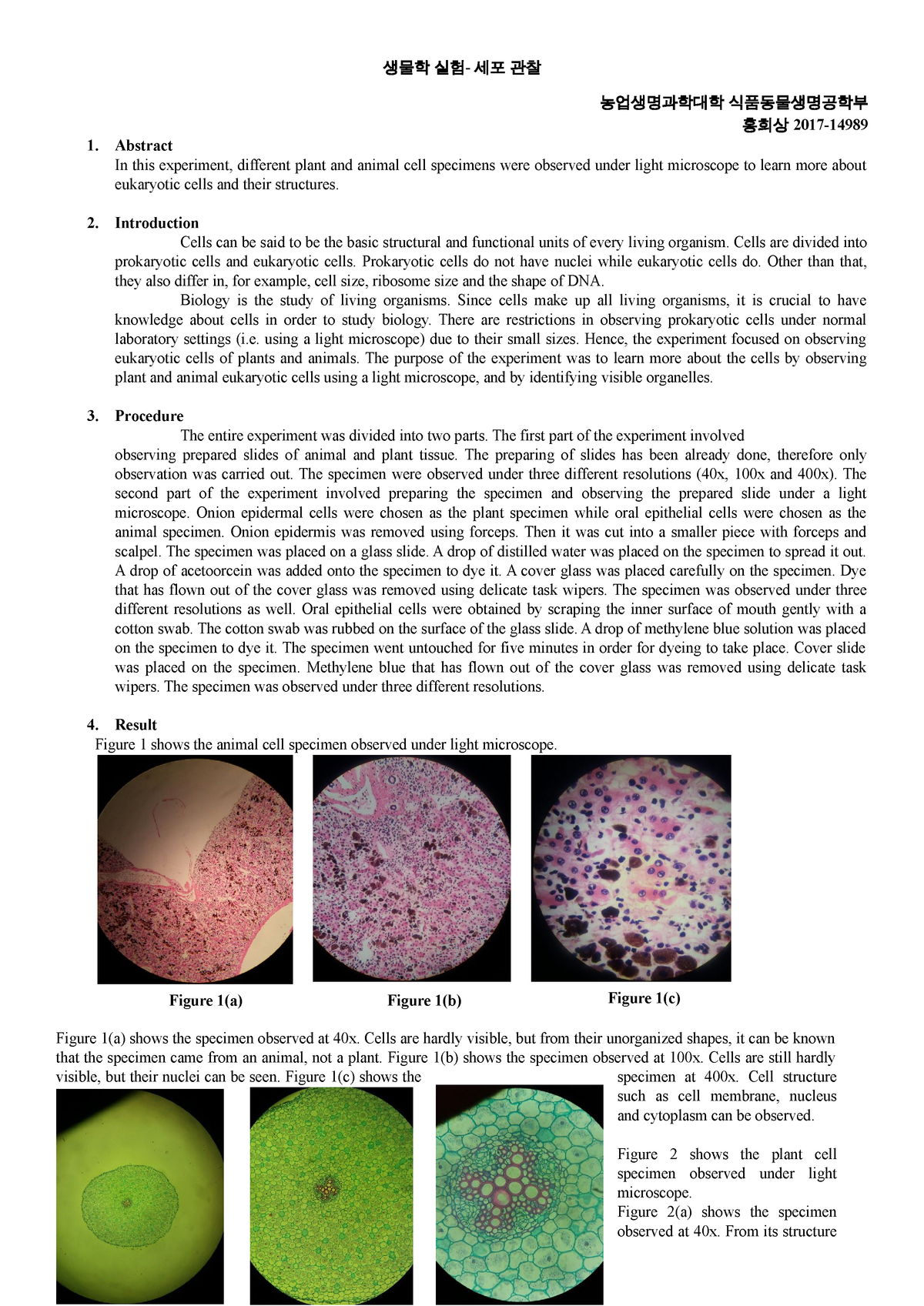
Animal Cell As Seen Under Light Microscope / diagram an animal cell as seen under light microscope ... - Under the light microscope, three parts can be seen in the animal cell:
byLaree Pacleb-
0
Animal Cell As Seen Under Light Microscope / diagram an animal cell as seen under light microscope ... - Under the light microscope, three parts can be seen in the animal cell:. Ppt eukaryotic cell seen under light microscope powerpoint. Electron microscopes use accelerated electron beams (as opposed to visible light in a light as for seeing electrons under any microscope in general, i would say we have come as close to it as plant cells look pretty much like animal cells except they have a cell wall and chloroplasts for. Each of these epithelial cells was examined under the microscope as students. (reproduced by permission of photo. Therefore we need to prepare the.
Light and electron microscopes allow us to see inside cells. Under a microscope, plant cells from the same source will have a uniform size and shape. Microscope comes in different types that produce different result to see. Resolving power is the ability to distinguish between separate things which are close to each other. It also has a very high resolving power.
Microscope image of plant vascular bundles under ... from i.pinimg.com Under a microscope, plant cells from the same source will have a. (ii)give your answers in b state the function of each of the following parts of a light microscope: Hydra under light microscopy stock photo download. Describe and compare the structure of a plant cell with an animal cell, as seen under a light microscope, limited to cell wall, nucleus, cytoplasm, chloroplasts, vacuoles and location of the cell membrane. Plant cells have cell walls, one large vacuole per cell, and chloroplasts, while animal cells will have a cell membrane only. Plant animal cells staining lab answers schoolworkhelper. Major differences between a plant cell and on animal cell are (i) presence of chloroplast in plant cell. (iii) presence of cell wall.
Present to a significant degree in animal cells) to generate contrast.
But in animals, there is a cell membrane n few small vacuoles. Animal cell cake of celliness: First seen with light microscopy 2. Under a microscope, plant cells from the same source will have a. Present to a significant degree in animal cells) to generate contrast. Animal cell under a microscope. Power house, provides cell with energy. The eyepiece and an objective lens. A generalised animal cell as observed under an electron microscope. Image:plant cell seen under electron microscope. Plant animal cells staining lab answers schoolworkhelper. The parts of a (palisade) plant cell that can be seen under a light microscope are:cell wallcell (surface) membranelarge (permanent) vacuolecytoplasmnucleuschloroplasts. (a)how is mitochondria adapted to its function?
Ppt eukaryotic cell seen under light microscope powerpoint. (ii) presence of large central vacuole in plant cell. A generalised animal cell as observed under an electron microscope. Keeps contents together, controls what goes in and out. Plant cells have cell walls, one large vacuole per cell, and chloroplasts, while animal cells will have a cell membrane only.
Animal Cells Under Light Microscope - Micropedia from d20ohkaloyme4g.cloudfront.net A generalised animal cell as observed under an electron microscope. (iii) presence of cell wall. Therefore we need to prepare the. Keeps contents together, controls what goes in and out. Light microscopy (the use of microscopes is called microscopy), in plant cells c. Central control, contains all information of chromosomes. Typical animal cell pinocytotic vesicle lysosome golgi vesicles golgi vesicles rough er (endoplasmic reticulum) smooth er (no ribosomes) cell (plasma) membrane… if you continue browsing the site, you agree to the use of cookies on this website. Animal cell cake of celliness:
Terms in this set (8).
Hey mate, here is your answer = under microscope, we have seen that plant cells have cell wall, large vacuoles. Each of these epithelial cells was examined under the microscope as students. Animal cell under a microscope. See our user agreement and privacy policy. Transports substances such as protein. The cell membrane is what controls the entry and exit of any substances that the cell needs to use or to dispose of. Given below is the diagram of a cell as seen under the microscope after having been placed in a solution Though we cannot see everything through the light microscope, some important organelles are visible and we can begin to see some structural differences between animal cells and plant animal cells under a light microscope. First seen with light microscopy 2. Cell membrane dr jastrow s electron microscopic atlas. They are very tiny than what human eyes can see in general. What was once unseeable can now be seen, touched, and eaten!cut yourself a wedge for dessert or snack on a nucleus, lyosome, or… Cell structures as seen under the light microscope.
1 4 1 observing plant and animal cells prac. Under the light microscope, three parts can be seen in the animal cell: Now the first thing to point out when looking at images under an electron microscope is the scale. Though we cannot see everything through the light microscope, some important organelles are visible and we can begin to see some structural differences between animal cells and plant animal cells under a light microscope. (iii) presence of cell wall.
Structure of Animal Cell and Plant Cell Under Microscope ... from i.pinimg.com Under a microscope, plant cells from the same source will have a uniform size and shape. Some features common to animal cells. Keeps contents together, controls what goes in and out. Magnification, however, is not the most important issue in microscopy. A generalised animal cell as observed under an electron microscope. Hey mate, here is your answer = under microscope, we have seen that plant cells have cell wall, large vacuoles. Ishita observed a slide of eukaryotic cell under electron microscope. Learn how to make an animal cell cake!
Ppt eukaryotic cell seen under light microscope powerpoint.
Hydra under light microscopy stock photo download. In most microscopes, there is a choice of. Therefore we need to prepare the. Under a microscope, plant cells from the same source will have a uniform size and shape. Differences between plant and animal cell. Ishita observed a slide of eukaryotic cell under electron microscope. Structures in an animal cell visible under a light microscope and an electron microscope. They are very tiny than what human eyes can see in general. (ii)give your answers in b state the function of each of the following parts of a light microscope: Typical animal cell pinocytotic vesicle lysosome golgi vesicles golgi vesicles rough er (endoplasmic reticulum) smooth er (no ribosomes) cell (plasma) membrane… if you continue browsing the site, you agree to the use of cookies on this website. See our user agreement and privacy policy. Animal cell under a microscope. Now the first thing to point out when looking at images under an electron microscope is the scale.